×
NOK
At the Department of Pathology at Rigshospitalet in Denmark, 8700 cryo-section examinations are performed annually, making the department one of the largest centers in Europe for this type of urgent examination. Cryo-section examinations are acute histological examinations in which the tissue is frozen to make sections for microscopic assessment. Cryo-sections are used partly for assessment of margin in connection with surgical cancer treatment and partly for determinating lesions and tumors - especially when cancer is suspected.
Over the past year, the Department of Pathology, in collaboration with University College Copenhagen, has worked hard to improve cryo-section diagnostics to make it even more specific and safe. An interdisciplinary project group has been established for the project, which is named: ACUT (Automated Cancer Analysis Under Twenty Minutes), in which Q-Stain plays a central role.
The project has received funding from the Danish Regions and the Health Cartel Development
and Research Fund and the interdisciplinary project group consists of: consultant Tina Agander, medical laboratory technologist supervisor Camilla Qvist, medical laboratory technologist Josefine Staldgaard, chief medical laboratory technologist Majbritt Wagner-Eckert, all from the Department of Pathology and associate professor Julie Smith from University College Copenhagen.
It is important for patients to get a quick clarification on their diagnosis. The Department of Pathology already has extensive experience with same-day assessment for head and neck patients in cancer patient pathways, where the patient is diagnosed and treated on the same day through cryo-section examinations.It helps to even out some of the inequality in the healthcare system.

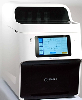
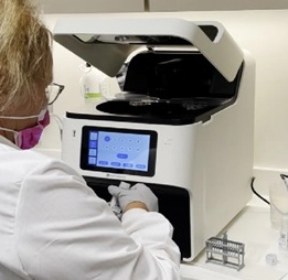
Q-Stain is implemented in the routine in connection with cryo-section examinations. It takes about 20 minutes to do immunohistochemical analyses (IHC) on the tissue, which should be added to the 20 minutes we already spend on making conventional cryo-sections. Within 45 minutes, it is possible for us to distinguish different types of cancer with much greater certainty. Our immune panel includes CD45, synatophysin, CKAE, CK5 and CK7 and eventually also Mart1, which can be used to distinguish carcinomas, and neuroendocrine tumors from lymphomas and melanomas. It can help us make even more reliable diagnoses – especially in cases where we are in doubt. In addition, it is important that a tumor with clear margins is removed during the surgical cancer treatment.
The diagnostic effect of IHC on cryo-sections can have direct treatment consequences for the patient, also referred to as the predictive value. For example, it can be difficult to type a tumor by conventional cryo-section examination. Thus, it can lead to a result, which can indicate a malignant tumor (cancer) awaiting further analysis and final diagnosis. In these cases, the IHC can optimally categorize the tumor as either: lymphoma (CD45+), melanoma (mart-1+), neuroendocrine tumor/carcinoma (synaptophysin+), or carcinoma (CK-AE+). This is important for severely ill patients who require emergency treatment, as the treatment depends on the type of tumor. Thus, for carcinomas, the first choice of treatment is radiation therapy, while for lymphomas it is chemotherapy.
ACUT plans to test and validate some of the predictive IHC markers that Novodiax is developing for Q-Stain. If we succeed in implementing relevant predictive markers in routine diagnostics, it will speed up clinicians’ ability to create a treatment plan early in the patient pathway. This will most likely improve the patient’s prognosis and save many hospital visits between diagnosis and start of treatment. This will probably also save resources in an already pressured healthcare system.
In collaboration with AH diagnostics, our department is the European reference laboratory for Q-Stain. We look forward to the collaboration and the continued development of the platform’s support for optimal patient pathways.

"Rigshospitalet in Denmark is at the forefront of raising the bar in cancer diagnostics. The Q-Stain is easy to use and fits into nearly any histology laboratory setting."
Helle Knakkergaard
Product Manager Pathology